In order to avoid, reduce and improve animal testing, the state of Baden-Württemberg founded a statewide network of universities on 8 February with the aim of improving animal welfare in research and teaching, and has made 3.8 million euros available for the purpose. The projects awarded funding also include the 3R-US network at the University of Stuttgart and the Robert Bosch Hospital in Stuttgart. The partners are developing an ex-vivo tumor tissue platform as a substitute for animal testing, and focus on molecular diagnostics, biomaterials and simulation.
Animal testing will remain an indispensable part of the repertoire of methods used in biomedical research for the foreseeable future. In the interests of animal protection as well as with an eye on the quality of the results, scientists are committed to continuously improving, reducing or avoiding animal testing – completely in keeping with the 3Rs principle (Replacement, Reduction, Refinement), which has been incorporated into German animal welfare law.
With the “3R-US” project coordinated by Prof. Monilola Olayioye from the Institute of Cell Biology and Immunology (IZI) at the University of Stuttgart, the teams at the IZI intend to continue implementing these standards together with researchers from the Institute of Biomaterials and Biomolecular Systems (IBBS) and oncologists from the Robert Bosch Hospital. Their aim is to make technology and methods of analysis available on a platform for cancer research which is based on the primary tumor tissue of human beings and which will gradually replace animal testing.
The group led by Monilola Olayioye is therefore researching the complex interactions between the signal networks of oncogenes, which promote the growth of tumors, and tumor suppressors, which inhibit it. These and other kinds of biophysical and biochemical mechanisms ensure that cancer cells cannot proliferate out of control. This makes it necessary to find suitable weak points for new substances which disable these mechanisms to latch onto. This way, the growth of the tumor can be stopped or the tumor can even be completely destroyed.
“If we want to test substances like these while carrying out as little animal testing as possible or at some point even none at all, we have to model the complex organism of a tumor as realistically as possible”, says Olayioye, speaking about the challenge she is faced with. Previous methods have only managed this to a very limited extent. The potential substance is fed directly into the cells during tests carried out in petri dishes with isolated cancer cells in a nutrient solution. This doesn’t correspond with the normal distribution processes within the body. Test systems whereby human tumor cells or tissues are implanted and grown in mice are approaching their limits however, since they don’t model the communication between cancer cells and immune cells in humans.
Ex vivo, de novo and in silico
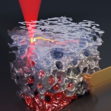
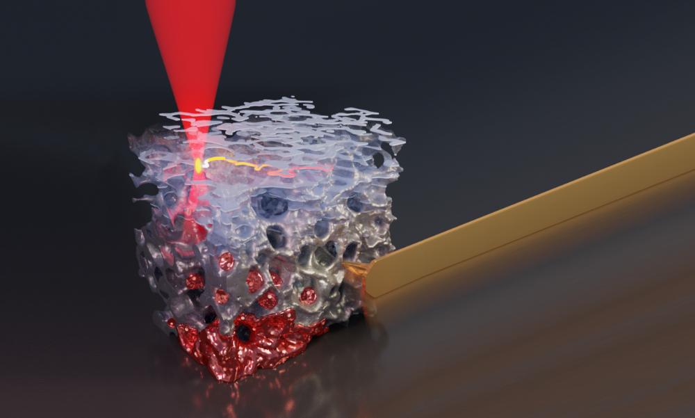
This is why researchers are looking for more ambitious systems, in which tumor cells can grow in three dimensions and can interact with other types of cells. 3R-US is based on three approaches: ex vivo, de novo and in silico. Ex vivo refers to a process whereby substances outside the organism are tested on tissue samples instead of individual cells. In order to obtain the different cell types and to understand the way they interact in the tissue, the team led by Prof. Walter Aulitzky at the Robert Bosch Hospital as well as experts from the affiliated Institute of Clinical Pharmacology has developed technology which enables human tissue samples, the so-called “slice cultures”, to be continuously supplied with nutrients in a controlled manner.
Since it is only possible to use this tissue once however, the 3R-US team focuses on reproducing the valuable material using so-called de-novo technology. To do this, it uses 3D printing techniques to “produce” similar structures to the tissue made from biomaterials and cells and cultivates them in a microfluidic system – tiny chambers which enable substances to be fed gradually. “By using these kinds of microsystem technologies we can assemble different biological structures, like using Lego bricks, so that they model the relevant tumor”, explains Michael Heymann, junior professor at the Institute of Biomaterials and Biomolecular Systems at the University of Stuttgart. The data which results from the ex-vivo and de-novo tests should also feed into creating and validating in-silico models. As a third component, more realistic forecasts for disseminating the substance should become possible by using models and simulations together with simulation experts.
“If we filter out the best drug candidates from ex-vivo culture systems at an early stage, we can significantly reduce the number of test animals used in the final preclinical tests”, says Prof. Roland Kontermann at the IZI. “De-novo and in-silico models offer a lot of potential for completely replacing animal testing one day.” Researchers want to share their knowledge about alternative test models with other partners from the 3Rs initiative. A 3R-US network spanning different institutions is set to be created over the next five years which should combine the knowledge of the university and the clinic, create synergies between expertise in biotechnology and medicine technology, and create a concept which can also be used to make students familiar with the topic at an early stage.
Expert Contact:
Prof. Monilola Olayioye, University of Stuttgart, Institute of Cell Biology and Immunology (IZI), Tel: +49 711 685-69301, E-Mail