
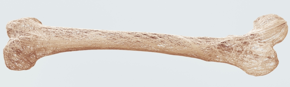
Asphalt, soil, the brain and bones all have one thing in common: they are all considered to be so-called porous media. Researchers at the University of Stuttgart are using the similarities between their physical behaviors to support medical research through simulations, for example by making progress in the understanding of multiple sclerosis and in the treatment of osteoporosis.
The brains of infants are often compared with a sponge, because the soak up information at this age. However, beyond this linguistic image, there is a much stronger physical similarity between the brain and a sponge – both are porous media. This term refers to structures that contain hollow spaces through which liquids can flow. Bones are also included among the porous media as are asphalt and garden soil. This explains why engineer Timo Koch is looking into medical questions, and also why he is completing his doctoral studies at the University of Stuttgart’s Institute of Modelling Hydraulic and Environmental Systems (IWS), where, actually, research is conducted into flows and transport processes in the subsurface terrestrial zone.
Whilst entirely different parameters, chronological and dimensional scales are of relevance to the medical issues that Koch is working on in collaboration with Professor Bernd Flemisch under the professorial auspices of Professor Rainer Helmig, ultimately these also involve fluid mechanics, i.e., providing a physical description of the propagation of a fluid throughout the porous structures of a given medium. In the case of the brain, the “porous medium” that Koch is studying, the cells and blood vessels form the structure in which the pores – the so-called intercellular spaces – are found.
Immune defense off the beaten track
Koch's scientific research is based on multiple sclerosis (MS) an autoimmune disorder in which the nerves become damaged. According to the German Multiple Sclerosis Society, it affects some 2.5 million people around the world, more than 200,000 in Germany alone. Most cases are diagnosed between the ages of 20 and 40. The initial symptoms are usually unspecific and include, numb patches on the skin, a tickly or numb feeling in the legs and blurred vision, and usually fade within a few days. The adverse effects often manifest themselves in surges, which can be alleviated for many sufferers through pharmaceutical drugs, which can also extend the period between subsequent surges.
However, the victims suffer motor impairments, which, in the worst case scenario, can result in their being confined to a wheelchair. They also suffer from attendant symptoms such as rapid exhaustion. The disease is incurable. Parts of the immune system fail to work correctly in MS patients and attack their own healthy bodies, resulting in damage to nerve cells and fibers. To diagnose and track the progress of the disease, medical practitioners use computed tomography, among other things, to scan the brain” Timo Koch explains. “The resulting images reveal areas where the blood-brain barrier has been compromised. The capillaries in these so-called lesions are far more porous than those in healthy brain tissue. Magnetic resonance imaging (MRI) can also be used for regular checks to see if new lesions have been formed and others have healed over, so that the doctors can adjust the treatment regime accordingly. In addition, a variant of this imaging process, the so-called perfusion MRI is also used in multiple sclerosis research, whereby the patient is injected with a contrast media, whose progress through the brain can be followed over time.
“The type and changes to the MRI signal enables us to determine where the contrast media is seeping out of the capillaries into the intercellular space”, Koch explains, and goes on to say that the immune system cells take the same path via these leaky areas to cause the unwelcome damage to the nerve cells and fiber. Koch's collaboration partners at the University of Bern’s ARTORG Center for Biomedical Engineering and the Neuroradiology Department at the University Hospital of Bern have discovered that the precise course of the MRI signal reveals whether a given lesion is still in the acute phase, or has started to heal or has completely healed over. Because of the low resolution of the MRI images, the Swiss research team is unable to determine from the data how much of the contrast agent has leaked out at a specific point. This is where Koch comes in, who simulates the propagation of the contrast medium on the computer.
To diagnose and track the progress of the disease, medical practitioners use computed tomography, among other things, to scan the brain”
PhD student Timo Koch
At first glance, the model that he uses for this looks nothing like a real brain. The researcher represents the capillaries as a row of tiny cylinders, which can cross over at various points. In the model, these capillaries cut through tiny, densely-packed cubes, which represent the intercellular space. Yet, the crucial factor is not what it looks like but rather how the characteristic physical equations – for such things as mass, pressure, velocity and concentration – change when the contrast medium seeps out through a leaky area in the capillary wall. “First”, Koch explains, “I have separate model equations for the capillary system and intercellular space respectively, which relate to the two different grids, i.e., to the different reference systems for the cubes and cylinders. I only amalgamate the two grids in the calculation itself”. One can envisage this so-called coupling as a process in which the two reference systems are reconciled or approximated to one another at the interfaces between the miniature cylinders and cubes.
Understanding flow processes within the brain
The result is a set of equations with around a million unknown variables, which Koch regards as “not that many” in terms of fluid mechanics per se. A single simulation for the propagation of the contrast medium takes about one to two minutes. “It takes several thousand iterations to be able to draw any reasonable conclusions – per pixel!”, he says and goes on to say that processes do exist that could be used to expedite the simulations. “But, that's not necessary, as, for the time being it's about fundamental questions of identifying the appropriate model parameters and calibration”, says the engineer.
The many iterations serve to estimate the parameters, i.e., the input variables, “whereby the results of the simulation are continually compared with MRI images of the patient's brain, to determine the best fit and, at the same time, most physically useful, meaningful parameters”, Koch explains. This process is done automatically, whereby the most relevant parameters are those that describe the physical properties of the materials. For instance, diffusion coefficients for the capillary walls can be found in the scientific literature or from the geometry of the capillaries. “A total of about ten parameters used in our model have an influence on the calculations”, says Koch. “The concentration profile with which the contrast medium moves through the capillaries is ultimately unknown, which means that you have to play through a reasonable range of values”. Finally, the researcher has to merge the flow model with the MRI images. Only then can useful conclusions be drawn about the development of the lesions. To this end, Koch also models the MRI imaging in a subordinated simulation. In this way, the researchers hope to gain a better understanding of the flow processes of an MS sufferer's brain than can be achieved with existing models.
We’re trying to use simulations to describe these processes.
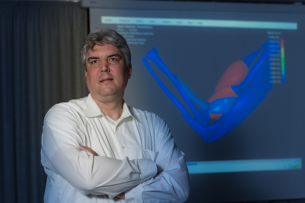
Oliver Röhrle, Professor for Continuum Biomechanics and Mechanobiology at the University of Stuttgart
Avoiding complications during treatment
Percutaneous vertebroplasty involves injecting a so-called bone cement into the vertebrae of patients suffering from osteoporosis. The treatment is minimally invasive: the surgeon gradually injects a few milliliters of the bone cement whilst continuously MRI images of the patient's brain, to determine the best fit and, at the same time, most physically useful, meaningful parameters”, Koch explains. This process is done automatically, whereby the most relevant parameters are those that describe the physical properties of the materials. For instance, diffusion coefficients for the capillary walls can be found in the scientific literature or from the geometry of the capillaries. “A total of about ten parameters used in our model have an influence on the calculations”, says Koch.
“The concentration profile with which the contrast medium moves through the capillaries is ultimately unknown, which means that you have to play through a reasonable range of values”. Finally, the researcher has to merge the flow model with the MRI images. Only then can useful conclusions be drawn about the development of the lesions. To this end, Koch also models the MRI imaging in a subordinated simulation. In this way, the researchers hope to gain a better understanding of the flow processes of an MS sufferer's brain than can be achieved with existing models. Special research for porous media The technical know-how about porous media available in Stuttgart is not just a reflection of Koch's research. In autumn 2017, the University received approval for a German Research Foundation (DFG) collaborative research center (CRC) focused on “boundary surface-driven multi-field processes in porous media”. Over 20 scientists from several of the University’s institutes participate in checking the results with x-ray images. “It's a standard treatment”, says Röhrle, “but, unfortunately, complications, such as bone cement leaking out of the vertebra, do occur from time-to-time. Moreover, the surgeon never knows how the bone cement will alter the mechanical behavior of the human musculoskeletal system”. Ultimately, the patient's vertebrae, ligaments, tendons and muscles will have adapted to the altered structure of the backbone. “Then there's the fact that, in the final analysis, the injected bone cement spreads differently in every patient”.
It's a standard treatment, but, unfortunately, complications, such as bone cement leaking out of the vertebra, do occur from time-to-time. Moreover, the surgeon never knows how the bone cement will alter the mechanical behavior of the human musculoskeletal system.
Oliver Röhrle, Professor for Continuum Biomechanics and Mechanobiology at the University of Stuttgart
From a fluid mechanics perspective, percutaneous vertebroplasty is a typical example for the processes in porous media. The injected bone cement sets within the vertebra, so that when it first enters the bone it causes a volume change and then, after setting results in a phase change from fluid to solid. “We’re trying to use simulations to describe these processes”, says Röhrle. And, at the same time, to take account of the properties of at least three materials – bone, bone marrow and bone cement. To validate the Stuttgart model, the scientists will be collaborating with the AO Research Institute Davos. “They have experimental laboratory set-ups there as well as the clinical problems that we require for our model development”, Röhrle explains. Only once the results of this first phase are available will the project participants be able to approach the question in which they are really interested: what exactly happens when a vertebra breaks or cracks?
Michael Vogel