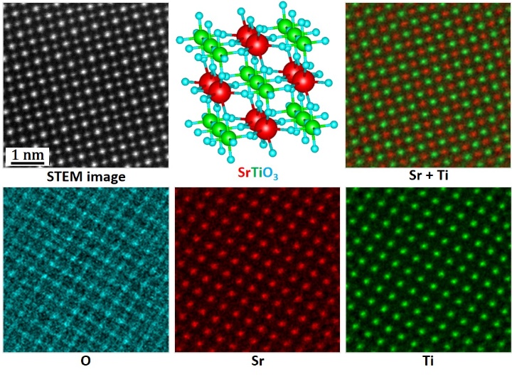
A new transmission electron microscope (TEM) at the University of Stuttgart that will be able to make visible tiny pores as small as 1 to 100 nanometers in size. The TEM Spectra-300 from the company Thermo Fisher is one of the most modern instruments worldwide for high-resolution examination of all kinds of materials. It is intended to advance the work of the Collaborative Research Center “Molecular Heterogeneous Catalysis in Confined Geometries” (SFB 1333), but also to be used in battery research and biology, for example.
The SFB 1333 (spokesperson Prof. Michael Buchmeiser) aims to accelerate chemical reactions and to produce particular products as effectively and specifically as in nature. This is to be achieved by means of novel catalysts that use defined, directing cavities as a reaction environment. “With the microscope, we want to find out whether the systems were produced successfully, we want to test their function and investigate how to improve them further,” says Prof. Guido Schmitz and Robert Lawitzki from the Institute for Materials Science, who will be operating the microscope at the University of Stuttgart. “Because the systems are only a few atoms in size and also very sensitive, a microscope with unique equipment is required to study them.”
The TEM allows the scientists to work with accelerating voltages in the range of 30 to 300 kilovolts, so that they can examine a wide range of specimens and achieve a very high resolution. One of the TEM’s features is the so-called probe Cs-corrector, which is located in front of the specimen in the illumination beam path and thus promises subatomic resolution below 0.05 nanometers in raster mode. An enclosure protects the microscope from acoustic vibrations and climatic influences, which ensures stable operation during high-resolution examinations.

Special features enable the examination of particularly sensitive specimens
Since the catalyst systems must not be damaged during the examination with the electron beam, the instrument is equipped with extremely sensitive detectors that can detect and evaluate signals even at low beam intensity. This includes a so-called direct-electron camera, a fast 4kx4k CMOS camera as well as other detectors for spectroscopic imaging. For particularly sensitive specimens, for example in cell biology, the instrument is also equipped with a cryo-transfer holder. Here, the specimens are cooled with liquid nitrogen and are thus more stable against beam damage.
Acquisition within the framework of the AMICA network
The TEM was acquired within the framework of the interdisciplinary network “Advanced Materials Innovation and Characterization” (AMICA) at the University of Stuttgart and will be set up in the Center for Electron Microscopy at the Materials Testing Institute (MPA). As a central facility, it will be used by 12 working groups from eight institutes, with more to follow. For example, the TEM can be used to perform in-situ experiments on novel solid-state batteries and look at what is needed at the atomic level to further increase the charging efficiency of these batteries. In biological research, the new microscope can broaden our understanding of proteins, tissues, and cells and to find out which mechanisms trigger certain processes or interactions at the nanoscopic level. Among other things, the researchers want to study the functioning of biological structures such as coronaviruses.
With the sum of EUR 4.2 million, the acquisition of the transmission electron microscope is a significant investment by the university in research infrastructure, which is supported by the German Research Foundation (DFG) with EUR 2.1 million within the framework of large-scale equipment funding. The installation, which required major spatial modifications at the MPA, will begin in April 2021. The microscope is expected to be handed over to the researchers in September 2021.
Expert Contact:
Robert Lawitzki, University of Stuttgart, , Institute for Materials Science, Chair of Materials Physics, Phone +49 711 685 61911, E-Mail


