A new sensor made from diamond is small and sensitive enough to detect the magnetic field of
nuclear spins in a volume as small as few nanometers. This is reported independently by two
research teams in the latest issue of "Science". One team involves groups from the universities of
Stuttgart and Bochum /Germany with additional partners, the other one is located at the IBM
research center Almaden / California. This breakthrough could lead to the development of a magnetic
resonance imaging (MRI) scanner operating at the nanoscale.
Such a device can be thought of as a powerful microscope, able to acquire three-dimensional
images of single molecules. It could trigger a revolution in biology, medicine and material science
alike.
Both groups employ a nitrogen-vacancy center in diamond as an atomic-scale sensor for magnetic
fields. This center is a red fluorescing color defect, responsable for the red color of some
gemstone diamonds. Using microwaves and laser light, a single center can be used as a magnetic
field sensor. Single centers can be embedded few atomic layers below the diamond surface by
shooting nitrogen ions into an otherwise pure diamond, a technique pioneered at the RUBION, the
central ion beam and nuclear facility of the University of Bochum. Such a shallow defect center can
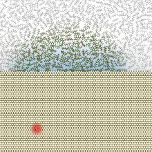
measure the magnetic field of molecules on the surface of a diamond.
A successful demonstration of such a measurement is the core result of both reports. Both
groups have used a single NV center to detect the magnetic field of nuclei in oil and plastic
coatings placed on the diamond surface. Below the coatings, they detected an oscillating magnetic
field at a specific frequency, as it is characteristic for hydrogen nuclei. This oscillating field
is also used by conventional magnetic resonance imaging scanners to generate an image, for instance
in hospitals. While these machines cannot reveal details smaller than a millimeter, the new
technique can detect nuclear spin signals from a million-fold smaller volume, only few nanometers
in size. This corresponds to a single biological molecule, for example an antibody.
So far, the detection of signals from such small samples has required elaborate machines,
working in high vacuum and close to absolute zero temperature. In contrast, the new technique works
at room temperature and merely consists of an industrial diamond in a microscope.
Operating in a low magnetic field, it is technically even simpler than clinical
scanners.
The next big challenge will be imaging, rather than detection, using the new sensor. In both
studies, the color center was fixed in the diamond lattice. While this rigid setup is sufficient to
measure the magnetic field signal it cannot acquire an image. In a way, the current result is only
the first pixel of a nanoscale MRI image. To acquire the entire image, the color center would have
to be moved over the sample by a nanopositioning apparatus such as an atomic force microscope.
Since this additional step appears realistic, the three-dimensional imaging of molecules might be
achievable in the near future.
This goal is vigorously pursued by many laboratories spread over the entire world. It can be
seen as a consequence of this fact that the current breakthrough was achieved simultaneously in two
labs.
Thanks to this global effort, the next big step might follow soon.
Contact:
Dr. Friedemann Reinhard, Universität Stuttgart,3. Physikalisches Institut, , Tel. 0711/685-65228, E-Mail: f.reinhard (at) physik.uni-stuttgart.de
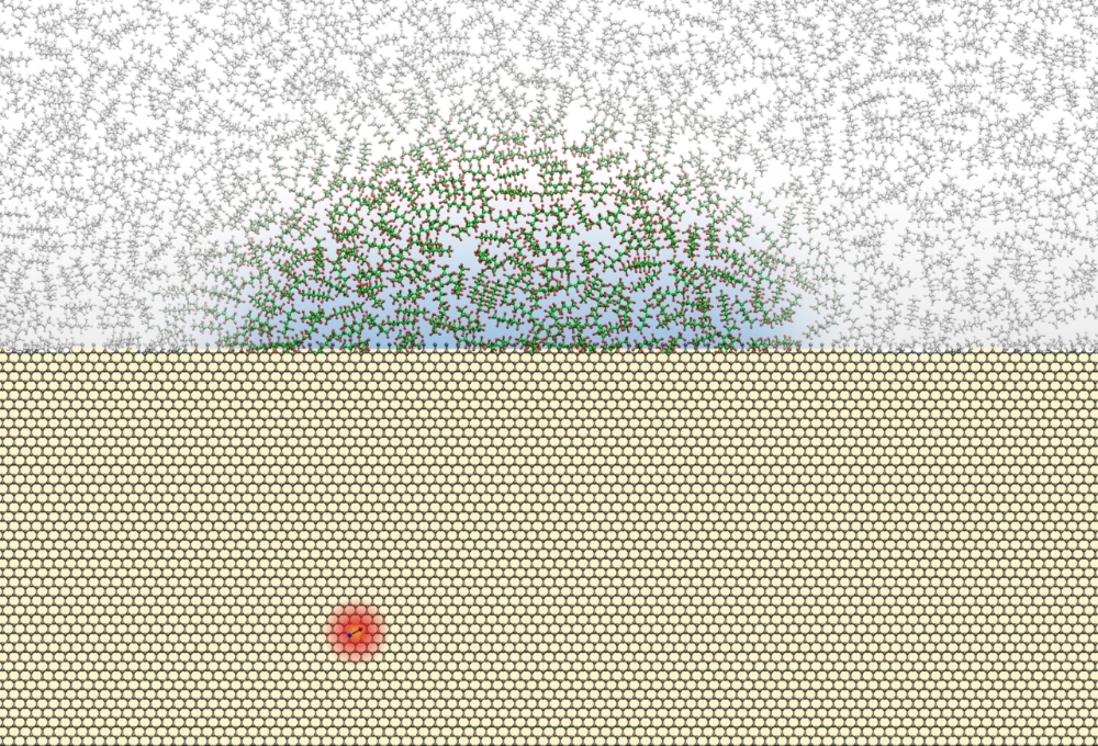
Visualisation of the experiment to detect the magnetic field of nuclear spins