
At first, it all sounds a bit unreal: Scientists are attempting to peer inside the body through the intact skin – without the aid of x-rays or magnetic fields. The process is called imaging with new scattering media, and medicine would be the primary beneficiary. The fact that the research community has been gripped by a downright spirit of pioneering optimism is also evident in a research project at the University of Stuttgart’s Institute of Applied Optics (ITO).
You would have to be Superman, who, as is well known, could look through any material except lead. Compared with this, the desiderata of medical practitioners seem rather modest: they would be satisfied with the ability to see through tissues, such as skin, which would enable them to identify changes to organs, cells or blood vessels without the need for surgical procedures. But we do already have a few of Superman’s powers at our disposal: There are x-rays, computed tomography and magnetic resonance imaging (MRI) with which, for example, bones or organs can be examined. However, the resolution of these methods is rather poor and requires exposure to radiation or strong magnetic fields. If, on the other hand, doctors could work with visible light, the results would be far more useful, because it significantly increases the achievable resolution, and because light interacts with many molecules within the body, which would provide additional information. To some extent, this is already possible today. For example, optical coherence tomography (OCT) makes it possible to look a few millimeters deep under the surface of the retina, which facilitates the early recognition of certain eye diseases.
However, the light only has to penetrate the transparent eyeball to do this: Skin represents a more formidable barrier, being between one and a half and three millimeters thick and apparently impenetrable by visible light. However, that is, in fact, not true as physicist Stephan Ludwig knows. Ludwig, a research assistant at the ITO, explains why the skin appears to be opaque in the following manner: “Rather than being absorbed by the skin”, he says, “the majority of ambient light that falls on it is scattered, in other words, reflected back in multiple directions”. For his doctoral studies, Ludwig is looking into the potential ramifications of this fact for new microscopes, still to be developed, which would enable doctors to see through skin.
Optical component with skin properties
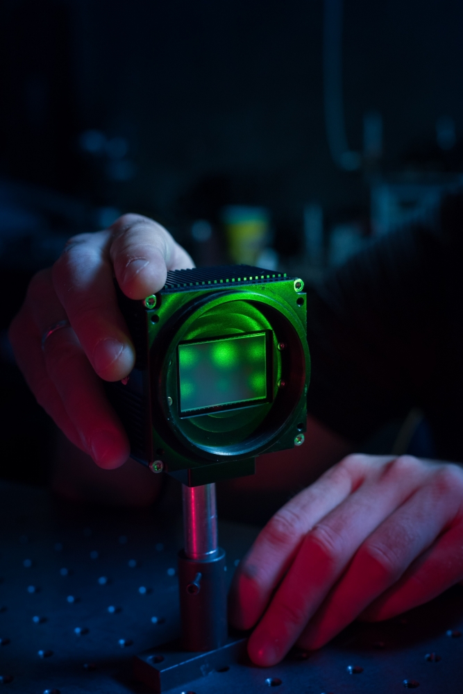
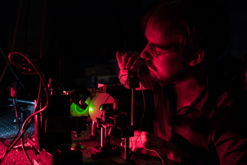
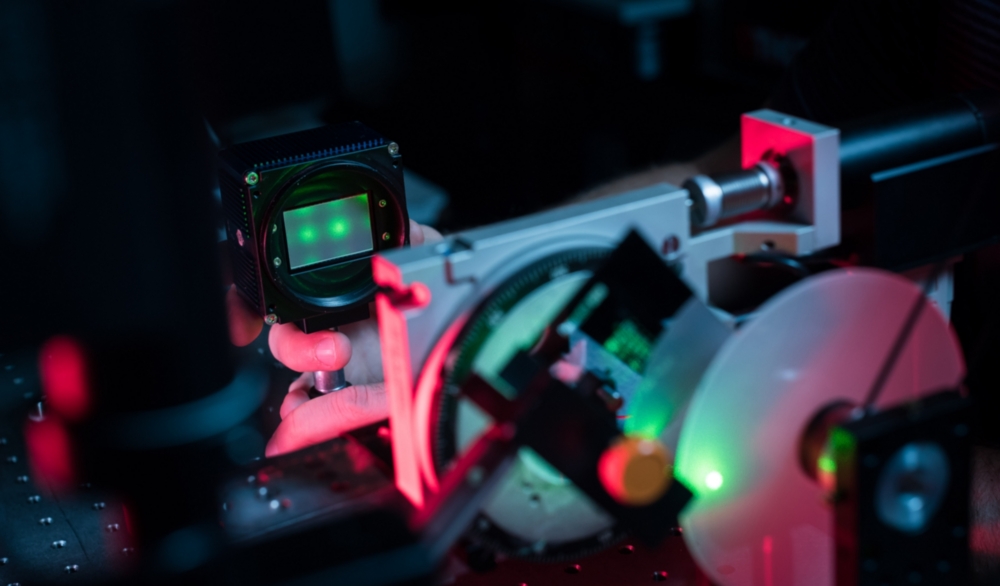
Until now, Ludwig has been undertaking fundamental research in the context of a project funded by the German Research Foundation, so there is currently no complete device standing in his laboratory. Instead, one finds all the technology that will be important for a so-called diffusion disc microscope in future. A simple test structure with lines placed at varying distances apart serves as the object to be examined. “But we’ve also already imaged onion cells, wood and plankton”, the physicist adds.
An optical microscope consists of a lens, a short distance in front of which the object to be examined is placed, and another lens with which the magnified image of the object can be viewed, or a camera, which can photograph the magnified image. The ITO researchers have exchanged the lens with a comparatively cheap diffusion disc – a glass disc roughened on one side – which can be bought for just 20 euro. This represents human skin, whose optical properties are similar to those of the human skin. From a physical perspective, this diffusion disc works as a lens, because it too produces an optical image. But, whilst a lens produces a sharp image of the object to be examined, the diffusion disc produces a pretty poor, out-of-focus image, just as skin would do. “But”, Ludwig interjects immediately,“in principle, this image still contains all the information about the object to be examined”. And this information can be distilled out with some computational effort.
That gives us an image of the simplest object there is – a single point of light
Stephan Ludwig, University of Stuttgart
Thus, taking a picture of something, such as a piece of wood, with the diffusion disc microscope involves two steps. First, a point light source serves as the object, whose characteristic light pattern created by diffusion disc is captured by the camera. “That gives us an image of the simplest object there is – a single point of light”, Ludwig explains. In the second step, the piece of wood is photographed through the diffusion disc, which is illuminated for this with light of the same color as the point light source. At first glance, this image also looks pretty chaotic. “But, because we know from the image of the point of light how the diffusion disc alters the image, we can also reconstruct the image of the piece of wood using mathematics.
Resolution in the millimeter range
It is true that the camera has not captured all of the scattered light, so that some information is, in fact, lost. Nevertheless, the quality of the image reconstructed in this way is astonishing. With his diffusion disc microscope, Ludwig is able to achieve a 40 x magnification and, therefore, can resolve structures down to the millimeter range. In addition, the diffusion disc microscope has other advantages over its conventional counterpart apart from a cheap lens. For example, the scientists can change the desired magnification level simply by varying the distances between the camera, diffusion disc and lens; in traditional microscopes the lens has to be changed for this. Another benefit is that the principle of the process can be transferred to other wavelength ranges from infrared to x-ray.

Yet, there are some downsides: in principle, the smaller the magnification level and the smaller the captured image, the better the image quality. Till now the ITO researchers have limited themselves to monochrome photographs. “But color images are our goal”, says Ludwig. Moreover, for an application that would actually go beneath the skin, for example, there is still no specific concept for how the object to be examined could be illuminated. “We might be successful with fluorescent nano particles injected into the body”, says the physicist, explaining one possible approach. “Ultimately”, and Ludwig makes no bones about this, “researchers still don't know the limits of this method”. However, imaging by means of scattering media has only really taken off experimentally in the last decade. “But, there has been incredible progress”, Ludwig finds. And even if peering through the skin turns out to be much too costly and complex for too small an added information yield, the relevant research could result in so many findings that it inspires applications outside of the medical field, such as for driverless navigation in foggy conditions, security checks or identifying paint delamination in works of art. Superman sends his regards.
Michael Vogel